Retinal Tears and Retinal Detachment
The vitreous gel in the middle of our eye changes over time. Some of it will form clumps which we see as floaters. The gel is also attached to the retina at the back of the eye. The gel will usually pull away from the retina and sometimes may develop flashes of light as this occurs. This process is called a posterior vitreous detachment.
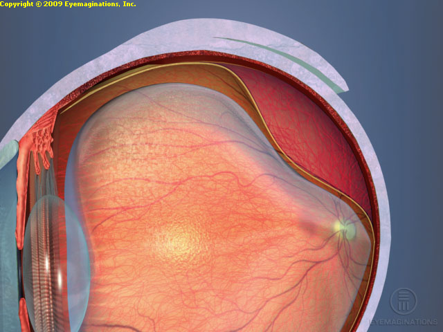
Usually, the vitreous gel pulls away from the retina cleanly and does not cause any problems. Sometimes the gel pulls strongly and tears a piece of retina. The retina is like wallpaper in the back wall of the eye. If there is a tear in the retina, fluid can go underneath the retina and cause a retinal detachment. This can cause vision loss and requires surgery.
If a retinal tear is seen before it becomes a retinal detachment, laser treatment can be applied to spot weld around the tear to lower the risk of developing retinal detachment by over 90%. As such, it is very important that any new flashing lights or floaters are evaluated with a dilated eye exam.
If a retinal detachment develops, then this requires surgery. The surgical techniques include a scleral buckle where a silicone band is placed around the outside of the eye, the fluid is drained with a small needle and freezing treatment called cryopexy is applied to the tear to cause a scar to attach the retina. Another surgical treatment is a vitrectomy where instruments are placed into the eye to assist in flattening the retina. This may require the use of a gas bubble and require positioning after surgery in a certain position to assist the retina in flattening.